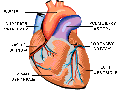
HeartThe heart is the organ that helps supply blood and oxygen to all parts of the body. It is divided by a partition or septum into two halves, and the halves are in turn divided into four chambers. The heart is situated within the chest cavity and surrounded by a fluid filled sac called the pericardium. This amazing muscle produces electrical impulses that cause the heart to contract, pumping blood throughout the body. The heart and the circulatory system together form the cardiovascular system.
Brain
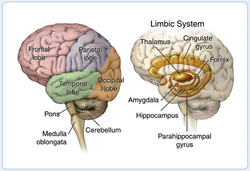
The forebrain is responsible for a variety of functions including receiving and processing sensory information, thinking, perceiving, producing and understanding language, and controlling motor function. There are two major divisions of forebrain: the diencephalon and the telencephalon. The diencephalon contains structures such as the thalamus and hypothalamus which are responsible for such functions as motor control, relaying sensory information, and controlling autonomic functions. The telencephalon contains the largest part of the brain, the cerebrum. Most of the actual information processing in the brain takes place in the cerebral cortex.
The midbrain and the hindbrain together make up the brainstem. The midbrain is the portion of the brainstem that connects the hindbrain and the forebrain. This region of the brain is involved in auditory and visual responses as well as motor function.
The hindbrain extends from the spinal cord and is composed of the metencephalon andmyelencephalon. The metencephalon contains structures such as the pons and cerebellum. These regions assists in maintaining balance and equilibrium, movement coordination, and the conduction of sensory information. The myelencephalon is composed of the medulla oblongata which is responsible for controlling such autonomic functions as breathing, heart rate, and digestion.
Lungs
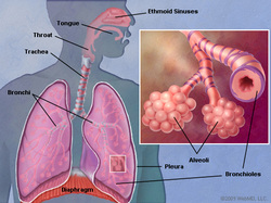
The lungs are a pair of spongy, air-filled organs located on either side of the chest (thorax). The trachea (windpipe) conducts inhaled air into the lungs through its tubular branches, called bronchi. The bronchi then divide into smaller and smaller branches (bronchioles), finally becoming microscopic.
The bronchioles eventually end in clusters of microscopic air sacs called alveoli. In the alveoli, oxygen from the air is absorbed into the blood. Carbon dioxide, a waste product of metabolism, travels from the blood to the alveoli, where it can be exhaled. Between the alveoli is a thin layer of cells called the interstitium, which contains blood vessels and cells that help support the alveoli.
The lungs are covered by a thin tissue layer called the pleura. The same kind of thin tissue lines the inside of the chest cavity -- also called pleura. A thin layer of fluid acts as a lubricant allowing the lungs to slip smoothly as they expand and contract with each breath
Liver
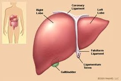
The liver is a large, meaty organ that sits on the right side of the belly. Weighing about 3 pounds, the liver is reddish-brown in color and feels rubbery to the touch. Normally you can't feel the liver, because it's protected by the rib cage.
The liver has two large sections, called the right and the left lobes. The gallbladder sits under the liver, along with parts of the pancreas and intestines. The liver and these organs work together to digest, absorb, and process food.
The liver's main job is to filter the blood coming from the digestive tract, before passing it to the rest of the body. The liver also detoxifies chemicals and metabolizes drugs. As it does so, the liver secretes bile that ends up back in the intestines. The liver also makes proteins important for blood clotting and other functions.
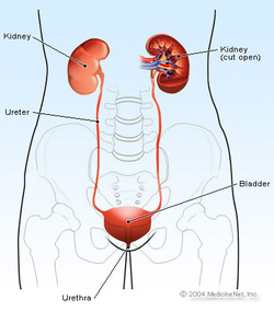
Kidneys
The kidneys play key roles in body function, not only by filtering the blood and getting rid of waste products, but also by balancing levels of electrolyte levels in the body, controlling blood pressure, and stimulating the production of red blood cells.
The kidneys are located in the abdomen toward the back, normally one on each side of the spine. They get their blood supply through the renal arteries directly from the aorta and send blood back to the heart via the renal veins to the vena cava. (The term "renal" is derived from the Latin name for kidney.)
The kidneys have the ability to monitor the amount of body fluid, the concentrations of electrolytes like sodium and potassium, and the acid-base balance of the body. They filter waste products of body metabolism, like urea from protein metabolism and uric acid from DNA breakdown. Two waste products in the blood can be measured: blood urea nitrogen (BUN) and creatinine (Cr).
When blood flows to the kidney, sensors within the kidney decide how much water to excrete as urine, along with what concentration of electrolytes. For example, if a person is dehydrated from exercise or from an illness, the kidneys will hold onto as much water as possible and the urine becomes very concentrated. When adequate water is present in the body, the urine is much more dilute, and the urine becomes clear. This system is controlled by renin, a hormone produced in the kidney that is part of the fluid and blood pressure regulation systems of the body.
Kidneys are also the source of erythropoietin in the body, a hormone that stimulates the bone marrow to make red blood cells. Special cells in the kidney monitor the oxygen concentration in blood. If oxygen levels fall, erythropoietin levels rise and the body starts to manufacture more red blood cells.
After the kidneys filter blood, the urine is excreted through the ureter, a thin tube that connects it to the bladder. It is then stored in the bladder awaiting urination, when the bladder sends the urine out of the body through the urethra.
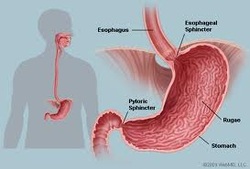
StomachThe stomach is an expanded section of the digestive tube between the esophagus and small intestine. Its characteristic shape is shown, along with terms used to describe the major regions of the stomach. The right side of the stomach is called the greater curvature and the left the lesser curvature. The most distaland narrow section of the stomach is termed the pylorus - as food is liquefied in the stomach it passes through the pyloric canal into the small intestine.The wall of the stomach is structurally similar to other parts of the digestive tube, with the exception that the stomach has an extra oblique layer of smooth muscle inside the circular layer, which aids in performance of complex grinding motions.
In the empty state, the stomach is contracted and its mucosa and submucosa are thrown up into distinct folds called rugae; when distended with food, the rugae are "ironed out" and flat.
In the empty state, the stomach is contracted and its mucosa and submucosa are thrown up into distinct folds called rugae; when distended with food, the rugae are "ironed out" and flat.
 RSS Feed