Heart Attack (myocardial infarction)
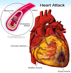
A heart attack (also known as a myocardial infarction) is the death of heart muscle from the sudden blockage of a coronary artery by a blood clot. Coronary arteries are blood vessels that supply the heart muscle with blood and oxygen. Blockage of a coronary artery deprives the heart muscle of blood and oxygen,causing injury to the heart muscle. Injury to the heart muscle causes chest pain and chest pressure sensation. If blood flow is not restored to the heart muscle within 20 to 40 minutes, irreversible death of the heart muscle will begin to occur. Muscle continues to die for six to eight hours at which time the heart attack usually is "complete." The dead heart muscle is eventually replaced by scar tissue.
Approximately one million Americans suffer a heart attack each year. Four hundred thousand of them die as a result of their heart attack.
What causes a heart attack?
Atherosclerosis
Atherosclerosis is a gradual process by which plaques (collections) of cholesterol are deposited in the walls of arteries. Cholesterol plaques cause hardening of the arterial walls and narrowing of the inner channel (lumen) of the artery. Arteries that are narrowed by atherosclerosis cannot deliver enough blood to maintain normal function of the parts of the body they supply. For example, atherosclerosis of the arteries in the legs causes reduced blood flow to the legs. Reduced blood flow to the legs can lead to pain in the legs while walking or exercising, leg ulcers, or a delay in the healing of wounds to the legs. Atherosclerosis of the arteries that furnish blood to the brain can lead to vascular dementia(mental deterioration due to gradual death of brain tissue over many years) or stroke(sudden death of brain tissue).
In many people, atherosclerosis can remain silent (causing no symptoms or health problems) for years or decades. Atherosclerosis can begin as early as the teenage years, but symptoms or health problems usually do not arise until later in adulthood when the arterial narrowing becomes severe. Smoking cigarettes, high blood pressure, elevated cholesterol, and diabetes mellitus can accelerate atherosclerosis and lead to the earlier onset of symptoms and complications, particularly in those people who have a family history of early atherosclerosis.
Coronary atherosclerosis (or coronary artery disease) refers to the atherosclerosis that causes hardening and narrowing of the coronary arteries. Diseases caused by the reduced blood supply to the heart muscle from coronary atherosclerosis are called coronary heart diseases (CHD). Coronary heart diseases include heart attacks, sudden unexpected death, chest pain (angina), abnormal heart rhythms, and heart failure due to weakening of the heart muscle.
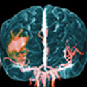
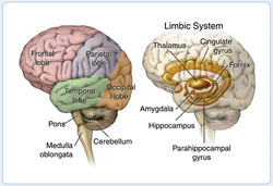
CVA (Stroke)A stroke (cerebrovascular accident, CVA, cerebral vascular accident or brain attack) occurs when a part of the brain is damaged or destroyed because it is deprived of blood.
There are 2 main types of strokes: ischaemic stroke and haemorrhagic stroke.
Ischaemic strokeIschaemic stroke is the most common type of stroke and is caused by a blockage of the blood vessels supplying the brain. This may be due to ‘hardening’ and narrowing of the arteries (atherosclerosis) or by a blood clot blocking a blood vessel.
One type of ischaemic stroke is a thrombotic stroke. This is caused by a blood clot (thrombus) in one of the arteries of the head or neck, which severely reduces the blood flow. The thrombus may be a result of a build-up of fatty deposits (plaques) in the blood vessels.
Another type of ischaemic stroke is an embolic stroke (or cerebral embolism), caused when a blood clot that forms elsewhere in the body (for example, the chambers of the heart) travels through the circulatory system to the brain. The travelling clot is called an embolus.
Haemorrhagic stroke. The most severe type of stroke is a haemorrhagic stroke. It occurs when a blood vessel in the brain bursts, allowing blood to leak and cause damage to an area of the brain. There are 2 types: subarachnoid haemorrhage, which occurs in the space around the brain; and an intracerebral haemorrhage, the more common type, which involves bleeding within the brain tissue itself.
The symptoms of a stroke usually appear suddenly. Initially the person may feel sick, and look pale and very unwell. They may complain of a sudden headache. They may have sudden numbness in their face or limbs, particularly down one side of their body. They may appear confused and have trouble talking or understanding what is being said to them. They may have vision problems, and trouble walking or keeping their balance. Sometimes a seizure (fit) or loss of consciousness occurs.
Depending on what function the damaged part of the brain had, a person may lose one or more of the following functions:
Symptoms occur rapidly and usually last a short time, from a few minutes to a couple of hours. Like a stroke, the symptoms will vary depending on which part of the brain is affected.
The warning signs
However, mini strokes should not be ignored as people who have had a temporary stroke are much more likely to have a stroke than people of the same age and sex who have not had a temporary stroke.
It is important that you see your doctor immediately when the warning signs of stroke occur. Your doctor will determine whether a stroke, a mini stroke or another medical condition with similar symptoms has occurred, such as a seizure or migraine.
There are 2 main types of strokes: ischaemic stroke and haemorrhagic stroke.
Ischaemic strokeIschaemic stroke is the most common type of stroke and is caused by a blockage of the blood vessels supplying the brain. This may be due to ‘hardening’ and narrowing of the arteries (atherosclerosis) or by a blood clot blocking a blood vessel.
One type of ischaemic stroke is a thrombotic stroke. This is caused by a blood clot (thrombus) in one of the arteries of the head or neck, which severely reduces the blood flow. The thrombus may be a result of a build-up of fatty deposits (plaques) in the blood vessels.
Another type of ischaemic stroke is an embolic stroke (or cerebral embolism), caused when a blood clot that forms elsewhere in the body (for example, the chambers of the heart) travels through the circulatory system to the brain. The travelling clot is called an embolus.
Haemorrhagic stroke. The most severe type of stroke is a haemorrhagic stroke. It occurs when a blood vessel in the brain bursts, allowing blood to leak and cause damage to an area of the brain. There are 2 types: subarachnoid haemorrhage, which occurs in the space around the brain; and an intracerebral haemorrhage, the more common type, which involves bleeding within the brain tissue itself.
The symptoms of a stroke usually appear suddenly. Initially the person may feel sick, and look pale and very unwell. They may complain of a sudden headache. They may have sudden numbness in their face or limbs, particularly down one side of their body. They may appear confused and have trouble talking or understanding what is being said to them. They may have vision problems, and trouble walking or keeping their balance. Sometimes a seizure (fit) or loss of consciousness occurs.
Depending on what function the damaged part of the brain had, a person may lose one or more of the following functions:
- ability to perform movements — usually affecting one side of the body;
- speech;
- part of vision;
- co-ordination;
- balance;
- memory; and
- perception.
Symptoms occur rapidly and usually last a short time, from a few minutes to a couple of hours. Like a stroke, the symptoms will vary depending on which part of the brain is affected.
The warning signs
- Sudden weakness or numbness of the face, arm and leg on one side of the body.
- Loss of speech, or difficulty talking.
- Dimness or loss of vision.
- Unexplained dizziness, especially when associated with any of the above signs.
- Unsteadiness or sudden falls.
- Headache (usually severe and of sudden onset).
- Confusion.
However, mini strokes should not be ignored as people who have had a temporary stroke are much more likely to have a stroke than people of the same age and sex who have not had a temporary stroke.
It is important that you see your doctor immediately when the warning signs of stroke occur. Your doctor will determine whether a stroke, a mini stroke or another medical condition with similar symptoms has occurred, such as a seizure or migraine.
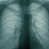
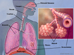
COPD (chronic obstructive pulmonary disease)
Chronic obstructive pulmonary disease (COPD) is one of the most common lung diseases. It makes it difficult to breathe. There are two main forms of COPD:
CausesSmoking is the leading cause of COPD. The more a person smokes, the more likely that person will develop COPD. However, some people smoke for years and never get COPD.
In rare cases, nonsmokers who lack a protein called alpha-1 antitrypsin can develop emphysema.
Other risk factors for COPD are:
Persons with COPD MUST stop smoking. This is the best way to slow down the lung damage.
Chronic obstructive pulmonary disease (COPD) is one of the most common lung diseases. It makes it difficult to breathe. There are two main forms of COPD:
- Chronic bronchitis, which involves a long-term cough with mucus
- Emphysema, which involves destruction of the lungs over time
CausesSmoking is the leading cause of COPD. The more a person smokes, the more likely that person will develop COPD. However, some people smoke for years and never get COPD.
In rare cases, nonsmokers who lack a protein called alpha-1 antitrypsin can develop emphysema.
Other risk factors for COPD are:
- Exposure to certain gases or fumes in the workplace
- Exposure to heavy amounts of secondhand smoke and pollution
- Frequent use of cooking fire without proper ventilation
- Cough, with or without mucus
- Fatigue
- Many respiratory infections
- Shortness of breath (dyspnea) that gets worse with mild activity
- Trouble catching one's breath
- Wheezing
Persons with COPD MUST stop smoking. This is the best way to slow down the lung damage.
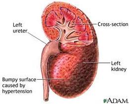
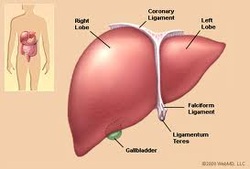
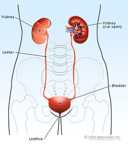
Kidney Failure (renal disease) Kidney disease is a general term that includes any disease, disorder or condition of the kidneys. The kidneys are vital internal organs located in the upper abdomen. Normally people have two bean-shaped kidneys, which form a part of the urinary tract in the genitourinary system.
Kidney disease is due to a variety of conditions that lead to kidney damage and deterioration of kidney function. Kidney disease can make it difficult or impossible for the kidneys to perform functions that are critical to life and your overall health including:
Kidney disease can be a serious or life-threatening condition because it can progress quickly and critically affect the ability of the kidneys to function normally. Seek immediate medical care (call 911)if you, or someone you are with, have symptoms of impaired kidney function, such as severe shortness of breath, bloody stools or urine, decrease in urinating or lack of urinating, or a change in consciousness or alertness. Seek immediate medical care (call 911) if you, or someone you are with, have overdosed on a drug or ingested a toxic substance.
SYMPTOMS Symptoms of kidney disease vary according to the underlying causes.
CAUSES Kidney disease can be caused by a wide variety of underlying diseases, disorders or conditions that lead to kidney damage, such as obstruction, infection, malignancy, inflammation, deformity, toxic ingestion, or a reduced blood supply to the kidneys.
TREATMENTS Treatment of kidney disease varies depending on the underlying disease, disorder or condition. The goals of treatment are to cure the underlying condition, prevent excessive fluid and waste from accumulating in the body, and stop or slow the progression of damage to the kidneys. Treatment also aims to minimize complications of kidney disease.
Kidney disease is due to a variety of conditions that lead to kidney damage and deterioration of kidney function. Kidney disease can make it difficult or impossible for the kidneys to perform functions that are critical to life and your overall health including:
- Filtering waste products and excess water and salts from the blood, which are then eliminated from the body through the ureters, bladder and urethra in the form of urine
- Producing certain hormones, such as renin, which helps regulate blood pressure
- Producing the active form of vitamin D (calcitrol)
- Regulating electrolytes and other vital substances, such as sodium, calcium and potassium
- Regulating the level and quality of fluid in the body
- Stimulating red blood cell production
- Acute renal failure is a condition in which there is damage and deterioration of kidney function that occurs suddenly, generally over a period of days. Acute renal failure can be caused by such conditions as shock, acute pyelonephritis, urinary tract obstruction, or ingestion of certain toxic substances.
- Chronic kidney disease is a condition in which there is damage and deterioration of kidney function that occurs over a long period of time, from months to years. Chronic kidney disease is generally caused by long-term diseases, such as diabetes and hypertension (high blood pressure).
Kidney disease can be a serious or life-threatening condition because it can progress quickly and critically affect the ability of the kidneys to function normally. Seek immediate medical care (call 911)if you, or someone you are with, have symptoms of impaired kidney function, such as severe shortness of breath, bloody stools or urine, decrease in urinating or lack of urinating, or a change in consciousness or alertness. Seek immediate medical care (call 911) if you, or someone you are with, have overdosed on a drug or ingested a toxic substance.
SYMPTOMS Symptoms of kidney disease vary according to the underlying causes.
CAUSES Kidney disease can be caused by a wide variety of underlying diseases, disorders or conditions that lead to kidney damage, such as obstruction, infection, malignancy, inflammation, deformity, toxic ingestion, or a reduced blood supply to the kidneys.
TREATMENTS Treatment of kidney disease varies depending on the underlying disease, disorder or condition. The goals of treatment are to cure the underlying condition, prevent excessive fluid and waste from accumulating in the body, and stop or slow the progression of damage to the kidneys. Treatment also aims to minimize complications of kidney disease.
 RSS Feed